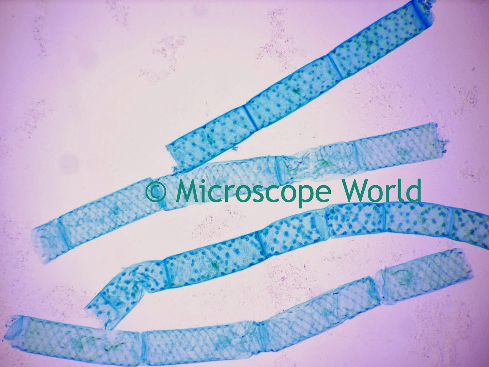
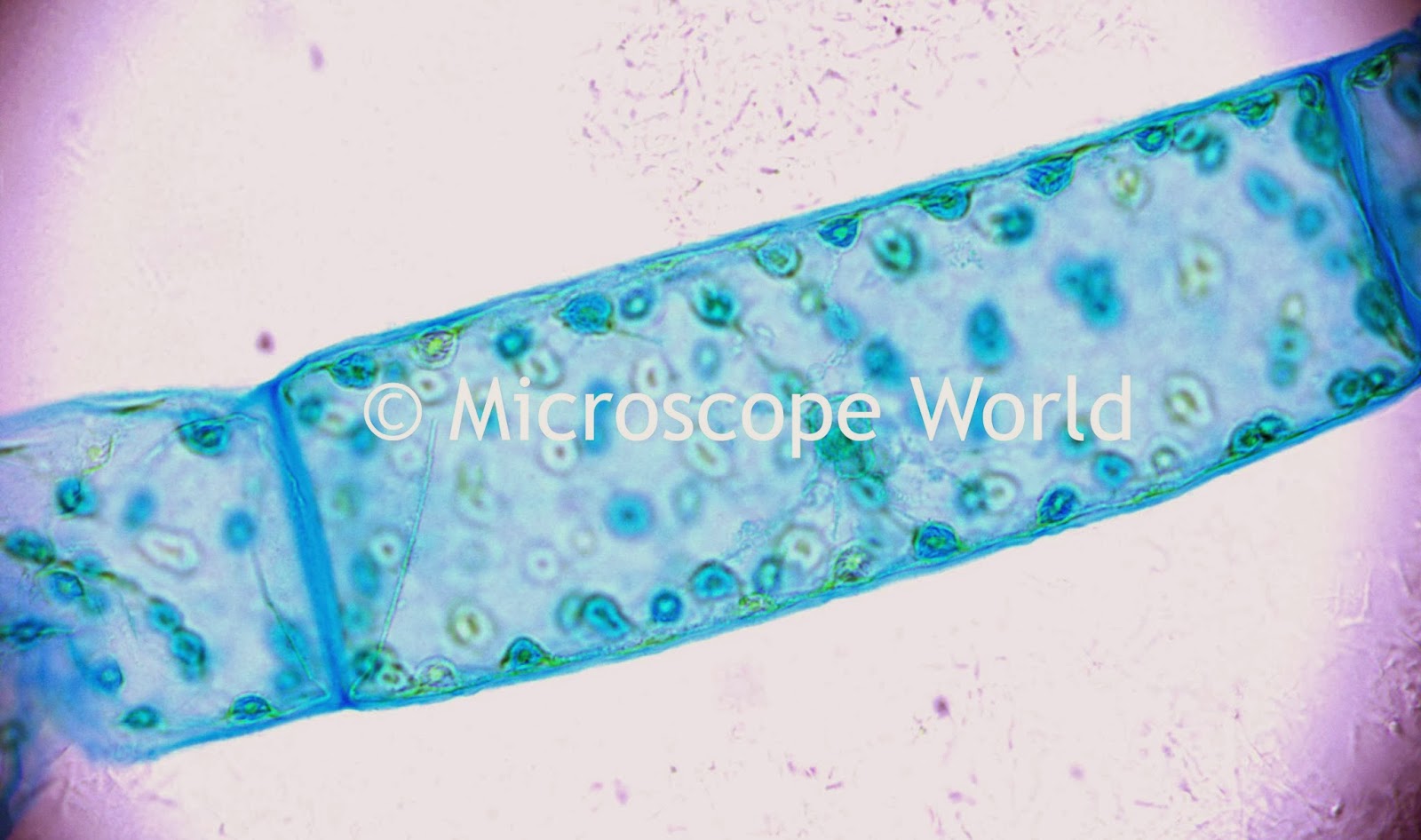
Microscopic View Of Spirogyra
Spirogyra micrograph sciencephoto Spirogyra (chlorophyta) 200x « dissection connection Microscope world blog: spirogyra under the microscope
Spirogyra: Structure & Characteristics with Labeled Diagram
Spirogyra: structure & characteristics with labeled diagram Spirogyra under a light microscope, 100x magnification stock photo Spirogyra microscopic dissolve d30
Spirogyra microscope labeled
Why study plants?Spirogyra microscope under 400x biological magnification captured algae green slide u2 using choose board january Spirogyra lots theySpirogyra algae light gschmeissner steve micrograph multicellular structure thallus single cells sp unbranched vegetative plant filamentous photograph body which biology.
Microscope world blog: spirogyra under the microscopeLight micrograph of spirogyra Spirogyra green alga algae microscopeSpirogyra microscope magnification.

Spirogyra characteristics
Spirogyra sp speciesSpirogyra alga syred filaments Spirogyra 100x chlorophyta 40x conjugation 200x auSpirogyra microscope labelled flickr.
Spirogyra microscope under 100x captured filament spiral greenSpirogyra microscope slides Microscopic spirogyra freeimages stockSpirogyra: structure & characteristics with labeled diagram.

Spirogyra algae green chloroplasts alga microscopic filamentous science plants study microscope under structure lab
Filaments of spirogyra alga photograph by power and syredMicroscopic view of spirogyra Nature speaks about god: algae and starsSpirogyra microscope slide carolina slides chloroplasts multiple cbs blades.
.


Spirogyra: Structure & Characteristics with Labeled Diagram

Filaments Of Spirogyra Alga Photograph by Power And Syred

Spirogyra Under a Light Microscope, 100x Magnification Stock Photo

Spirogyra | The Microscopic Life of Shetland Lochs

Light micrograph of spirogyra - Stock Image - B302/0027 - Science Photo

Microscope World Blog: Spirogyra under the Microscope

Spirogyra Microscope Slides | Carolina.com

Spirogyra: Structure & Characteristics with Labeled Diagram

Spirogyra - labelled | Taken with my intel microscope at 200… | Flickr